Varus
In this circumstance, a extra anterior pores and skin incision, adopted by a formal arthrotomy, was carried out, as a concomitant lateral femoral condyle osteochondral allograft switch was carried out. Once the lateral femoral cortex is satisfactorily exposed, a meta-diaphyseal guidepin is inserted, starting on the lateral cortex, and positioned at an angle towards the medial femoral epicondyle. After the osteotomy is made, the scale of the allograft bone wedge insert relies on the amount of correction decided preoperatively. The allograft is placed quickly in a press fit fashion, whereas the general limb alignment is checked on intraoperative radiographs.
Our approach corresponds to this and the common HKA and MAD of our patients indicate a postoperatively centered, and never a new, lateralised, mechanical axis. This is defined by the completely different etiologies in our research group, including younger sufferers with out structural harm but with medial knee ache. For these patients the aiming point of the new mechanical axis is the medial intercondylar tubercle and for patient with grade IV medial cartilage degeneration the lateral one. DFO can reliably correct valgus mechanical alignment of the lower extremity, decrease ache, and improve perform in sufferers with lateral compartment illness. The osteotomy could be performed in a medial closing-wedge or lateral opening-wedge method.
Clinical Diagnostics And Imaging
The mediolateral diameter of the osteotomy site is measured intraoperatively by measuring the size of the two initial guidewires that are positioned from medial to lateral. The intraosseous lengths of the anterior and posterior guidewires are then averaged to supply the diameter reference for the chart.10 This wedge size ought to be used to guide placement of the second set of 2 guidewires that determine the wedge resection measurement. Using fluoroscopic guidance, a guide pin is positioned approximately 2 to 3 fingerbreadths proximal to the lateral epicondyle and aimed simply proximal to the medial epicondyle. This will determine the angle of the osteotomy made first with the oscillating noticed, and followed by osteotomes.
- Both medial closing-wedge and lateral opening-wedge osteotomies of the distal femur have been reported for correction of genu valgum.5 Patient-reported knee high quality of life is improved by either approach.6, 7, eight, 9 Advantages of each approach are detailed in Table 1.
- To request a price quote and/or surgical evaluation of merchandise, please add all products of interest to your cart by deciding on the respective product checkboxes within the “Quote” and/or “Surgical Evaluation” column of the “Products View” .
- This database will further our detailed understanding of osteotomy surgical procedure.
- Only the research by van der Woude et al. investigated the postoperative clinical consequence after a cDFO so far and reported a Lysholm score of 73 points and a pain level of 3 .
Among them, Salter-Harris type II is the most common, making up about half of progress plate fractures, whereas types IV, V, and VI (Rang’s type VI) are rare, accounting for only a few % . Havranek and Pesl reported patients treated for Rang’s kind VI physeal damage constituted 36 youngsters (zero.12%) of children with acute fractures with imply age was eleven.6 years. In the present case, we consider SH type VI perichondral ring damage was initially present as a result of the fracture was brought on by a valgus harm and hemorrhage around the peripheral construction of the growth plate and no obvious signs of a fracture have been noted on MRI. Many earlier authors have tried to quantify risk factors for the potential for a growth arrest occurring, together with intercourse, SH types , age, displacement, therapy, and ligamentous laxity . Premature closure of the expansion plate is recognized by the looks of a bony bridge within the fractured development plate. With respect to treatment, though elongation surgery of the affected limb is chosen for rising sufferers, lengthy-term observe-up is recommended in some circumstances as spontaneous correction in some instances have been reported.
A Dedicate Instrumentation For A Exact Surgical Procedure
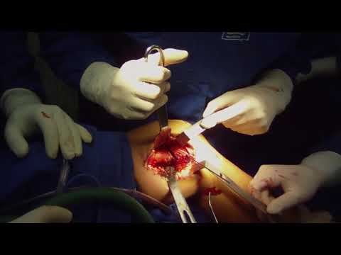
Only after the mechanical axis has been corrected will the plate be positioned and secured on the lateral femoral cortex. Although OA is extra prevalent in females,6 the literature is inconclusive as to the gender during which the procedure is most regularly performed.sixty seven In our research, 12 osteotomy sufferers have been male, whereas 14 had been feminine. After the osteotomy, the entire circumstances achieved a neutral anatomical alignment, with the valgus angle ranging from 0° to 1°. A, joint line marking, patella and surgical access; B, subvastus retractor placement; C, parallel pins with information plate placement; D, proximal a part of the osteotomy; E, placement of pins on the wedge slicing information to complete the osteotomy; F, plate placed after osteotomy.
A 2.zero-mm threaded K-wire is positioned in the anterior distal screw gap to provisionally fix the plate in place . The proximal aspect of the plate ought to be positioned parallel to the middle of the femoral shaft. Calibrated locking guides are screwed into the distal plate, and the posterior distal screw is drilled and positioned in locking fashion unicortically. The other 3 distal screws are then positioned in unicortical locking style to the metaphyseal section . A bicortical nonlocking screw is used to bring the plate right down to the diaphyseal femur and compress the osteotomy site .
The bones are secured in position with the help of metallic plates and screws. Dr. Wheeless enjoys and performs all kinds of orthopaedic surgical procedure however is renowned for his expertise in complete joint arthroplasty in addition to complex joint infections. He founded Orthopaedic Specialists of North Carolina in 2001 and practices at Franklin Regional Medical Center and Duke Raleigh Hospital. In general, the plates and screws which might be used to fix lengthy bone fractures are left in for a minimum of one year prior to having them taken out. This is because there is usually a higher rate of fracture after hardware elimination of plates and screws which are eliminated prior to at least one 12 months after their placement. Sharma L., Song J., Felson D.T., Cahue S., Shamiyeh E., Dunlop D.D. The function of knee alignment in illness development and functional decline in knee osteoarthritis.
There was no conversion to whole knee arthroplasty in a comply with-up of at least 5 years. One affected person had a superficial infection handled with cleaning and antibiotics, without having for the removing of the plaque and of the screws. The current study was evaluated and permitted by the Ethics and Research Committee of this institution beneath the quantity CAAE .9.0000.5505. FJ, PS, WF, DN, PC, and TPH contributed to the interpretation of the outcomes. BW corrected the manuscript and gave directional enter all through the examine.
Hospital For Special Surgery
The regular mechanical axis of the lower limb is defined as a line passing from the middle of the femoral head, by way of the center of the knee, and persevering with down to the center of the ankle. This differs from the anatomic axis of the lower limb, which follows a line from the middle of the femoral head, down the femoral shaft through the center of the tibia on the knee joint, to the middle of the ankle, as depicted in Figure 1. Abnormal lateral distal femoral angles are thought-about something lower than 84 levels. Standard radiographic evaluation features a bilateral standing full-size alignment view, bilateral weight-bearing anteroposterior views in full extension, bilateral weight-bearing posteroanterior tunnel views at 30 levels of flexion , lateral, and sunrise or Merchant views.
To describe the surgical strategy of distal closing-wedge femoral osteotomy and a instances sequence submitted to this method. This research evaluates radiological and clinical midterm consequence of re-alignment procedures in case of varus deformtities. The importance of comparing preoperative planning with actual postoperative alignment and the associated medical consequence is emphasised.